
On this page
At a glance
- A cone beam CT scan builds a 3D image of your jaw from a single, short scan.
- It shows bone height and width, the nerve canal, the sinus floor and root shapes that flat X-rays can't.
- It's used mainly before implants, grafts or certain surgeries, when the extra depth will change the plan.
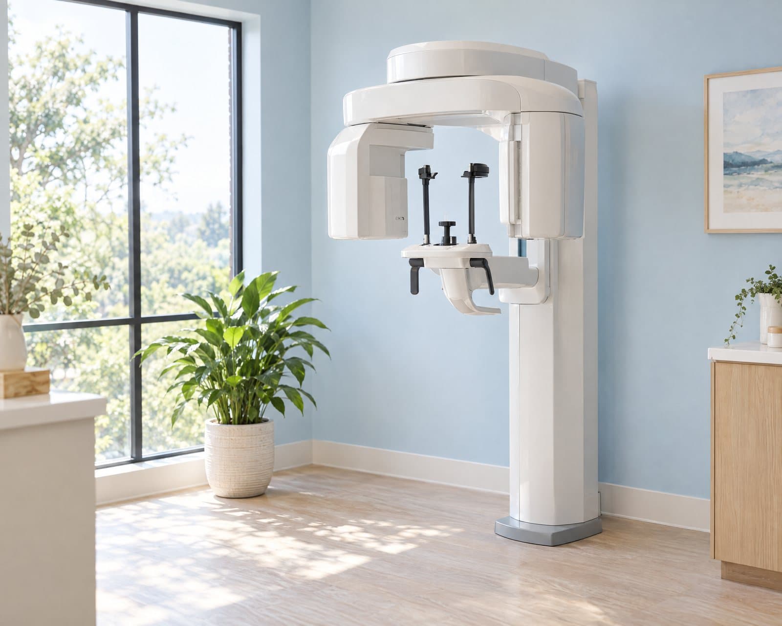
- The scan itself is quick, seated or standing, with no enclosed tube.
What is a cone beam CT scan?
A cone beam CT scan is a type of dental X-ray that captures a 3D image of your jaw, teeth and surrounding structures in one short pass. Unlike a hospital CT scan, it's built specifically for the mouth, jaws and face.
The machine circles your head once, and software assembles the images into a 3D model your periodontist can rotate and measure. That model is far more detailed than a flat X-ray, though it still uses X-ray energy to build it.
What does a 3D scan show that X-rays can't?
Regular dental X-rays are flat images, so they compress a 3D jaw into two dimensions. A cone beam CT scan keeps the third dimension, showing bone height and width, not just length and shading.
It also maps structures that matter for planning: the nerve canal in the lower jaw, the floor of the sinus above the upper back teeth, and the shape of tooth roots nearby.
That extra depth is what makes guided implant surgery possible: a plan built from the scan can guide exactly where an implant goes, before any incision is made.
| What differs | Regular dental X-rays | Cone beam CT |
|---|---|---|
| Views | Flat, two-dimensional images | A 3D model you can rotate and measure |
| What it shows best | Decay and bone level along the tooth | Bone height, width, nerves and sinus position |
| Radiation | Very low, for a single image | More than one flat X-ray, well under a hospital CT scan |
| Typical uses | Routine checkups and everyday care | Planning implants, grafts and some surgeries |
When is a cone beam CT scan used?
A 3D scan is common before dental implants, especially when bone height or width is in question. It's also used before some grafting procedures and surgeries, when a flat X-ray can't show the structures clearly enough.
It's often ordered before a sinus lift, which adds bone under the sinus in the upper back jaw, and before ridge augmentation, which rebuilds a thin ridge. In both cases, seeing the area in three dimensions changes how the procedure is planned.
What is a cone beam CT scan like?
You're seated or standing, not slid into an enclosed tube. A guard rotates around your head once while you stay still and often bite on a small positioning piece; the machine doesn't touch you.
The scan itself takes well under a minute, though the visit takes longer with positioning and any questions you'd like answered beforehand. There's little to prepare beyond removing metal jewelry or a removable retainer.
What are the risks of a cone beam CT scan?
A cone beam CT scan uses more radiation than a single dental X-ray, but much less than a hospital CT scan, and only the area being planned is scanned. It's used when that extra detail will actually change the plan, not as a routine step.
If you're pregnant, tell us beforehand, since imaging decisions are made with extra care in that case. Guided surgery built from the scan supports the plan; it doesn't replace a clinician's judgment during the procedure.
What affects the cost of a cone beam CT scan?
Cost depends on how much of the jaw needs to be scanned and why the scan is needed. A scan for one implant site costs less than a scan covering a full arch or both jaws.
See the exams and technology overview for how a 3D scan fits alongside other tools and tests.
- How much of the jaw or face needs to be captured
- Whether the scan is for planning implants, a graft or a surgery
- Whether guided surgery is planned from the images
- Whether your dental plan helps cover 3D scans
Care at Perio One
At Perio One in Lafayette, a cone beam CT scan is ordered only when the extra detail will change your plan, not as a routine step. Dr. Randal Comeaux, DDS, Diplomate of the American Board of Periodontology, reviews the images with you before deciding on next steps.
Every implant and graft plan here starts with an examination, and 3D cone beam CT planning is added when it brings real value, as it often does for implants. Findings are explained in plain language, and we coordinate with your general dentist throughout.
Referrals are welcome, and implants planned from the scan are followed in our periodontal maintenance program.
What to expect
- Referral or recommendation
Your periodontist recommends a scan when 3D detail will change how implants, a graft or a surgery is planned.
- Getting ready
Remove metal jewelry or a removable retainer; there's little else to prepare.
- Positioning
You're seated or standing, and a small guard rotates around your head once while you stay still.
- The scan
The capture itself takes well under a minute; the rest of the visit is positioning and any questions you have.
- Reviewing the images
Dr. Comeaux reviews the 3D model with you and explains what it means for your plan.
Common questions
What does a cone beam CT scan show?
It shows a 3D view of bone height and width, the nerve canal, the sinus floor and the shape of tooth roots. That depth helps plan implants, grafts and some surgeries more precisely than flat X-rays alone.
Is a cone beam CT scan safe?
It uses more radiation than a single dental X-ray but much less than a hospital CT scan, and only the area being planned is captured. It's recommended when the extra detail will actually change your plan.
Is a cone beam CT scan necessary?
Not for every visit. It's used selectively, mainly before implants, grafts or surgeries where the extra depth changes how the procedure is planned; simpler cases often don't need one.
How much radiation is in a dental cone beam CT scan?
It's more than a single flat dental X-ray and far less than a hospital CT scan, though the exact amount depends on the machine and the area captured. Only the region needed for your care is scanned.
What is guided implant surgery?
It's implant placement planned in advance from your 3D scan, so your periodontist knows the position and angle before starting. It supports the plan; the clinician's judgment still guides the procedure itself.
Sources
- Cleveland Clinic: Dental X-rays
- MedlinePlus: Dental x-rays
- American Academy of Periodontology: Dental implant procedures
This page is general education, not a diagnosis. Your periodontist will recommend care after an examination.